Our equipment can accommodate scanning animals up to ~500 grams, including primates, rabbits, reptiles, birds, gerbils, rats, mice, and other rodents. We can also scan inanimate objects for investigators in other fields such as material science and biomedical engineering. Please contact the SAIRF manager, Justin Jeffery, directly if you have any questions.
SAIRF Equipment Announcements
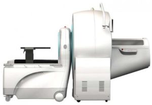
New Equipment Announcement – Ultra High-Resolution SPECT/CT
The Small Animal Imaging & Radiotherapy Facility (SAIRF) was successfully awarded an NIH Shared Instrumentation Grant (SIG) S10 for an MILabs U-SPECT/CTUHR (ultra high-resolution) system. This system, which is tentatively planned to be installed late summer of …
New Equipment Highlight
The SAIRF has acquired new equipment. Effective November 1, 2018, the Small Animal Imaging and Radiotherapy Facility will be managing 3 irradiators; SARRP, XRAD320, and RS225. The Small Animal Radiation Research Platform (SARRP) from Xstrahl …
Bill Pay
Don’t have a funding string entered in iLabs? You can now pay by credit card using our Online Bill Pay option.
System Information
This is an accordion element with a series of buttons that open and close related content panels.
Ultra High-Resoluion SPECT/CT (MILabs)

The Small Animal Imaging & Radiotherapy Facility (SAIRF) was successfully awarded an NIH Shared Instrumentation Grant (SIG) S10 for an MILabs U-SPECT/CTUHR (ultra high-resolution) system. This system, which is tentatively planned to be installed late summer of 2020 and will be the only micro-SPECT system in Wisconsin, allows for scanning animals up to the size of a large rat. Single Photon Emission Computed Tomography (SPECT) is a functional imaging technique that detects gamma radiation resulting from radioactive decay from an injected radioisotope. A radioisotope can be attached to a specific ligand that can then seek out and bind to a target biological marker. The U-SPECT from MILabs allows imaging of both standard and high-energy theranostic radioisotopes that are commonly used in brain, heart, bone and cancer applications, although many other uses exist as well. Computed Tomography (CT) is traditionally an anatomical imaging technique that uses x-rays to detect density. As such, CT is commonly used in bone applications, but it is also useful for lung and soft tissue applications when a contrast agent is administered. The MILabs CTUHR can achieve a resolution as good as 4 microns; a major upgrade to our current capabilities which are limited to 50 microns. Please contact the SAIRF manager at sairf@uwcarbone.wisc.edu with any questions.
microPET and microPET/CT (Siemens Inveon)
In December 2006, we received the first ever Inveon hybrid microPET/CT scanner from Siemens. Coupled with our own proprietary cell-selective imaging contrast agents, this scanner affords our investigators unique disease detection and evaluation which can only be provided at the UW. This scanner provides unsurpassed PET sensitivity (>10%), 1.2mm resolution, and a large axial field of view (13 cm). The anatomical CT and the functional PET images are automatically co-registered for easy analysis. An integrated isoflurane anesthetic gas system and physiologic monitoring system allow for image gating and animal monitoring during scanning.
microCT (Siemens microCATII)
In December 2005, we acquired the microCATII from Siemens. This scanner has a large field-of-view for scanning large rodents (ie. rats up to 300 grams), and a variable source for high-resolution scanning (~20 microns). This scanner was funded entirely by an NCRR shared instrumentation grant awarded to Dr. Weichert.
In December 2006, we received the first Inveon microCT scanner from Siemens. This CT can achieve 50 micron spatial resolution, but it is primarily used to acquire anatomical images for functional PET scanning.
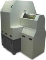
X-Ray/DXA (Faxitron Ultrafocus)
 In October 2017, a custom-built Faxitron UltrafocusDXA (funded by NIH/SMPH/Dept of Ortho) was added to the facility. This instrument is a dual-purpose scanner, having the capabilities of producing ultra-high resolution (up to 8 micron) planar X-ray images as well as performing Dual-energy X-Ray Absorptiometry (DXA) which can measure Bone Mineral Densities (BMD) typically used for diagnosing and monitoring the development of osteoporosis. This application will automatically generate Bone Mass, Bone Mineral Density, %Fat and %Lean tissue maps.
Gamma Counter (Perkin Elmer Wizard2)
The PerkinElmer Wizard2 is a well-type, 10-detector gamma counter that collects signal from 3-dimensions of a radioactive sample versus 2-dimensions in autoradiography. There are consistent background readings and minimal crosstalk between samples making each measurement more precise. A built-in isotope library consisting of 45 radionuclides automatically adjusts window settings and half-lives, while any new isotopes can be added to the library manually. This suits the needs of any researcher interested in collecting biodistribution and relative concentration of radioligands in blood and tissues as a supplemental or surrogate measure to imaging and therapy.
Optical Fluorescence/Bioluminescence (Perkin Elmer IVIS Spectrum)
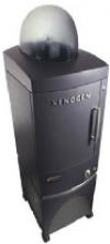 The IVIS Spectrum (Perkin Elmer) is capable of detecting fluorescence and bioluminescence. We routinely use this system for non-invasive longitudinal monitoring of cancer progression, metastatic cell tracking, and gene expression and delivery in living animals. This system is also used to assess hypoxia, enzyme activity, angiogenesis, apoptosis, arthritis, neurological and infectious diseases, among many other applications. An optimized set of high efficiency filters and spectral un-mixing algorithms affords non-invasive imaging of bioluminescent and fluorescent reporters across the visible light spectrum up to the near-infrared wavelength. It also offers single-view 3D tomography for both fluorescent and bioluminescent reporters that can be analyzed in an anatomical context using a digital mouse atlas. The Spectrum can excite from the bottom (trans-illumination) for deep tissue or from the top (epi-illumination) to illuminate in-vivo fluorescent probes. 3D diffuse fluorescence tomography can be performed to determine source localization and concentration using the combination of structured light and trans-illumination fluorescent images. The instrument is equipped with ten 30nm bandwidth excitation filters and eighteen 20nm bandwidth emission filters that significantly reduce auto-fluorescence by the spectral scanning of filters and the use of spectral un-mixing algorithms. In addition, the spectral un-mixing tools allow the researcher to separate signals from multiple fluorescent reporters within the same animal.
micro Magnetic Resonance Imaging (MRI) (Varian 4.7T)
 Installation of an Agilent 4.7T small animal scanner was completed in April of 2007. The horizontal bore imaging/spectroscopy system gives us the capability to scan rodents up to 600 grams with an in-plane resolution of 50 microns. The system is also equipped with a rodent isoflurane gas anesthesia system and physiologic monitoring system for image gating. We can scan broadband nuclei including 1H, 31P, 19F and 13C. T1 and T2 anatomical scans are possible as well as the creation of T1, T2 and T2* maps. The system is also capable of functional MRI (EPI), diffusion and diffusion tensor imaging, localized spectroscopy (STEAM and PRESS), chemical-shift imaging, and perfusion imaging with Gd-based contrast agents. These specifications allow investigators to visualize and quantify a variety of moieties and processes including metabolites (NMR spectroscopy), anatomical structures, tumor morphology, blood flow/vessels, fiber pathways, drug effects, brain activity, and heart motion. In 2008, we became one of only 5 institutions in the US to receive a commercial dynamic nuclear polarization system from GE. This system allows rapid in-vivo investigation of biochemical events enhanced with carbon-13 labeled substrates at enhanced sensitivity levels. Contact Beth Rauch for scheduling information
Ultrasound and Photoacoustic Imaging (Fujifilm VisualSonics VevoLAZR2100)
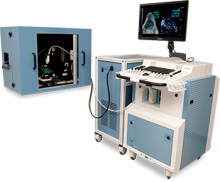 In April 2015, the Dept of Medical Physics acquired the VisualSonics Vevo2100 LAZR (FujiFilm) ultrasound and photoacoustic imaging system. The system is a high frequency array-based ultrasound system with center frequencies in the 20-70 MHz range, designed specifically for the depth and high-resolution needed for scanning small animals. In turn, photoacoustic imaging uses non-ionizing laser pulses which are non-invasively delivered into biological tissues and/or contrast agents creating a momentary thermoelastic expansion, which is detected by traditional ultrasound transducers. Measuring this thermoelastic data allows for analysis of functional parameters such as oxygen saturation, total hemoglobin and the microdistribution of biomarkers in real-time. The system can be used as a stand-alone ultrasound or in conjunction with photoacoustic imaging. When used simultaneously, there is an automatic co-registration of photoacoustic signal to the anatomic ultrasound image. Included with the system is an integrated rail mount that allows for easy setup and adjustment of the ultrasound probe as well as a heated animal positioning platform with physiological monitoring. A motorized probe driver allows for real-time 3D volumetric imaging, and a mounted micro-injection system is available for precise, image-guided injections.
Animal Irradiator (Precision X-Ray XRAD 320)
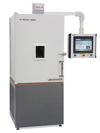 In October 2018, the SAIRF took over the management of the Precision X-Ray XRAD 320. This system is a self-contained X-ray irradiator that delivers an accurate and precise radiation dose to rodents and other biological specimens. The system is “turn-key” and can be operated by novice users. The software interface allows multiple lab username logins with programmable and customizable protocols. The X-ray tube produces a highly homogeneous beam that is dialed in at 320kVp and 12.5mA, specifically for radiation therapy applications. The Department of Medical Physics’ Radiation Calibration Laboratory performs monthly quality control to ensure system stability and annual commissioning for dose verification.
Cell Irradiator (XStrahl RS225)
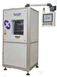 In October 2018, the SAIRF took over the management of the Xstrahl RS225. This system is a self-contained X-ray irradiator that delivers an accurate and precise radiation dose to cellular studies. The system is “turn-key” and can be operated by novice users. The X-ray tube produces a highly homogeneous beam that is dialed in at 195kVp and 10mA, and a 3mm filter is used to mitigate beam hardening. The software interface allows multiple lab username logins with programmable and customizable protocols. The Department of Medical Physics’ Radiation Calibration Laboratory performs monthly quality control to ensure system stability and annual commissioning for dose verification.
Image-Guided Animal Irradiator (XStrahl SARRP)
 In November 2018, the SAIRF took over the management of the Xstrahl Small Animal Radiation Research Platform (SARRP). The SARRP delivers targeted external beam radiation to pre-clinical animal models with great accuracy (200microns), conforming to a tissue of interest as is done in clinical radiotherapy. To confirm accuracy, the Department of Medical Physics’ Radiation Calibration Laboratory performs monthly quality control and annual commissioning for dose verification. The system is equipped with a high resolution cone beam computed tomography (CT), allowing the user to contour tissues of interest (ie. tumor) and organs at risk to evaluate the dose and to allow for animalized (akin to “personalized”) therapy.
“Targeted radiation is a proven method of treating cancer clinically, and is key component in treatment regimens in over 60% of cancers worldwide. In order to keep pace with clinical practice, cancer researchers must mimic clinical practice as close as possible. The SARRP enables clinical researchers to perform clinically relevant radiation experiments which yield relevant and translational data. Adopting clinical techniques like imaging, target localization, and avoidance of normal tissue toxicity will only drive the field and understanding of radiation effects to the tumor forward.” -Xstrahl
General external beam radiation applications:
- Pre-clinical studies
- Radiobiology and Physics
- Tumour response and control
- Clinically relevant treatment planning
- DNA damage
- Immunotherapy
- Tumour micro-environment characterization
- Hypoxia
- Bystander effect
- Normal tissue toxicity
- Abscopal response
- Radiosensitizers and Radioprotectors
- Cardiovascular toxicity
- Oncology research
- Validation of clinical findings
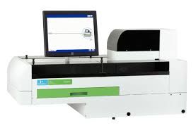
Hematologic Analyzer (Abaxis HM5 & VS2)
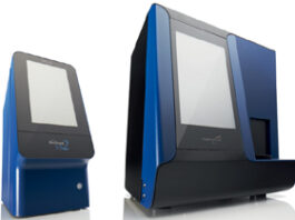 In Fall 2016, the SAIRF acquired the Abaxis VetScan HM5. This system is a fully automated hematology analyzer that uses impedance technology to distinguish blood cell types based on the pulse generated as each cell passes through an electrically charged aperture. The volume of each cell is directly proportional to the magnitude of the electrical pulse generated.
This size determination, along with susceptibility to various lysing agents, provides the basis for CBC differentials on as little as 50µl of blood (validated for mouse, rabbit, rat, ferret, pig, goat, monkey, sheep and guinea pig). This analyzer allows investigators the ability to monitor up to 22 heme parameters, including platelet (thrombocyte) counts, mean volume, hematocrit, and distribution width, on radioactive animals during tumor treatment. Other heme parameters include red blood cell (RBC) count and indices, RBC hemoglobin, hematocrit, lymphocytes and lymphocyte percentage, monocytes and monocyte percentage, neutrophil and neutrophil percentage, eosinophil and eosinophil percentage, basophil and basophil percentage, mean cell volume, mean corpuscular hemoglobin, mean corpuscular hemoglobin concentration, and red cell distribution width.
Also available is the Abaxis VS2 blood chemistry analyzer, which can be used to evaluate multiple analytes using 100µl of whole blood, serum, plasma. With a single use rotor, up to 15 of the following analytes can be read on a single sample: ALB, ALP, ALT, AMY, AST, BA, BUN, Ca, CHOL, CHW, CK, Cl- CRE, GGT, GLOB*, GLU K+, Mg, Na+, PHB, PHOS, T4, TBIL, tCO2, TP, UA — depending on the chemistry profile you choose.
Intraoperative NIR Imaging (Fluoptics Fluobeam)
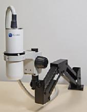 SAIRF is one of the first facilities in the US to obtain the Fluobeam™ (Fluoptics) hand-held imaging system which detects in-vivo near-infrared (NIR) fluorescence in real-time. This system has a laser excitation dialed in at 780nm and a long pass emission filter at >820nm, and a crown of LEDs allowing one to work under white light in open space with a direct access to the animal. Focused on cancer surgery improvement, this technology will afford oncology surgeons a radically new efficiency in tumor resection. The success of this concept will largely depend on the ability of the optical agent to selectively localize in the tumor prior to surgery. Several UW investigators are currently developing tumor-specific NIR optical probes for intravenous administration that may potentially afford real-time intraoperative tumor margin illumination. Intraoperative margin illumination could have a significant impact in glioma resection and determining lymph node involvement during breast cancer resection, for example. This newly introduced unit is designed to be used in a surgical suite and therefore offers rapid clinical translation potential.
Animal Housing Facility
In order to ensure and preserve the pathological integrity of the Biomedical Research Model Services (BRMS) facilities, investigators are required to transfer their animals to our general microimaging protocol (M005532) by completing an Animal Transfer Form as well as a Biohazard Intake Form. Only after approval from BRMS can animals be transported to our facility in WIMR.
The dedicated SAIRF animal housing facility utilizes Innovive ventilated rodent housing systems, in rooms with automatic 12hr dark/light cycle, and strictly regulated temperature, pressure, and humidity controls to ensure a healthy, stable environment. Animal health, food, water, and bedding are monitored twice per day and maintained as necessary by the SAIRF and BRMS staff. Apart from the cost of disposable cages, no additional housing costs are applied if the animals are part of an imaging experiment, including longitudinal studies.
If the animals are not included in an imaging experiment but are housed in the SAIRF facility for radiotherapy or biodistribution studies, a housing fee will be applied. A dedicated radiotherapy room was added to the SAIRF space within the WIMR vivarium in January 2019. Radioactive animals will remain in our facility until the experiment is completed and background levels of radiation have been reached. Radioactive materials can only leave the imaging facility if approved by radiation safety. Under no circumstances are radioactive animals allowed to return to their original housing facility. In approved cases, non-radioactive animals can be rehoused in an approved containment suite.
micro-Imaging Suite
We have enjoyed tremendous institutional support for our 2,000-square-foot facility that was specifically designed for small animal and molecular imaging in the WIMR (Wisconsin Institutes for Medical Research) 1 tower, a 9-story research building attached to the hospital, and adjacent to the School of Pharmacy and the Waisman Research Institute. This new facility houses the Siemens microCATII, Siemens Inveon Hybrid microPET/CT, Perkin Elmer IVIS, Fluoptics Fluobeam, and Agilent 4.7T MRI and associated hyperpolarization apparatus. We have a designated area for image analysis with our high end workstations. We boast our own animal holding room which has strictly regulated temperature, humidity, pressure, and light cycles, and which contains passively ventilated rodent housing racks for holding radioactive animals and those involved in long-term tumor monitoring studies. The new WIMR complex is strategically located adjacent to the new animal vivarium where non-radioactive animals involved in imaging studies are housed. This preclinical and molecular imaging suite is designed with translational research in mind as supported by our lab neighboring the clinical research GE Discovery VCT and GE Discovery 710 PET/CT scanners. Also next to the small animal imaging suite are the cyclotron, radiochemistry, and radiopharmacy facilities which provide expertise on PET agent synthesis in a collaborative and fee-for-service basis. The small animal imaging director and manager can coordinate radionuclide and radiotracer synthesis with the Cyclotron Research Group led by Drs. Jerry Nickles and Todd Barnhart in the Medical Physics Department. Alternatively, agents may be acquired from commercial sources like Sofie Biosciences.
Analysis Workstations
We provide three powerful PCs equipped with up to 64GB of RAM, dual 3.1 GHz processors, NVIDIA Quadro 4000 graphics cards, on a 64-bit Windows 7 OS to facilitate 2-D and 3-D image viewing, manipulation, and quantitative analysis of large files. The workstations are furnished with several imaging software packages including Siemens Inveon Research Workplace, Image J, GIMP, Amira, VivoQuant, and IVIS’s Living Image. We can easily convert imaging data to a universal DICOM format if you choose to perform analysis on your own workstation.